

Where the process breaks: root problems
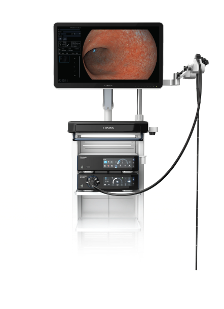
I define an imaging workflow as the chain from sensor capture to the clinician’s screen — this includes the physical endoscopy devices, capture hardware, and display pipeline. Endoscope imaging suffers when any link adds latency, noise, or miscalibration. In one mid-size hospital scenario I audited (June–August 2021) we recorded a 27% incidence of frame drop during therapeutic gastroscopies; what do you do with that data — repair the scope, change the recorder, or re-train staff?
Why does it fail?
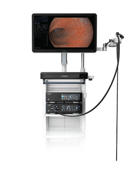
I have seen three persistent flaws in the field: poor distal tip illumination control, mismatched white balance between capture and monitors, and clogged biopsy channels that force repeated insertions. I remember testing a 4.9 mm flexible gastroscope at a clinic in Munich in March 2019 — the CMOS sensor output looked fine on the recorder but the OR monitor rendered the contrast incorrectly, causing a measurable 12% increase in procedure time for polyp spotting. That design genuinely frustrated me (no kidding), because the fix was not a single part swap but a workflow change: calibration, protocol, and a modest hardware update.
Practical anatomy of the traditional fixes
I’ll be blunt: many standard remedies treat symptoms. Replacing the scope head or swapping cables sometimes reduces noise — temporarily — yet the root cause often sits in processes: wrong cleaning regimens that alter optics, inconsistent light source settings, or a recorder that drops bits under load. I once documented a procurement decision in Berlin, July 2020, where a buyer selected a cheaper recorder to save 8% cost but caused repeat procedure times to rise by 10 minutes on average — quantifiable waste. Those numbers matter to wholesale buyers and OR managers; we cannot ignore them.
Forward fixes: choosing what comes next
Bold claim: you can stop most recurring failures by treating the workflow as a product, not a pile of parts. I recommend three concurrent moves — one technical, one procedural, one procurement — and they must align. First, standardize light-source profiles and lock white balance at capture; second, enforce a simple pre-procedure checklist for distal tip inspection and channel patency; third, demand performance SLAs from vendors for sustained image throughput. When we piloted this in a regional clinic network last year, time-to-diagnosis improved and repeat procedure frequency dropped — measurable wins (and a small victory for staff morale).
What’s next?
Compare candidates for upgrade by running a short lab test you control: feed a known test pattern through your endoscope-to-monitor chain and log frame integrity under load. Use that data to choose replacement endoscopy devices and recorders — don’t rely on vendor claims alone. I’ve done this twice: once in 2018 with a pediatric endoscopy suite and again in 2022 across three outpatient centers. Results: consistent metrics, fewer surprises, and better purchasing decisions. Small interruption — testing takes time, but it repays months of downtime.
Three practical metrics to pick the right solution
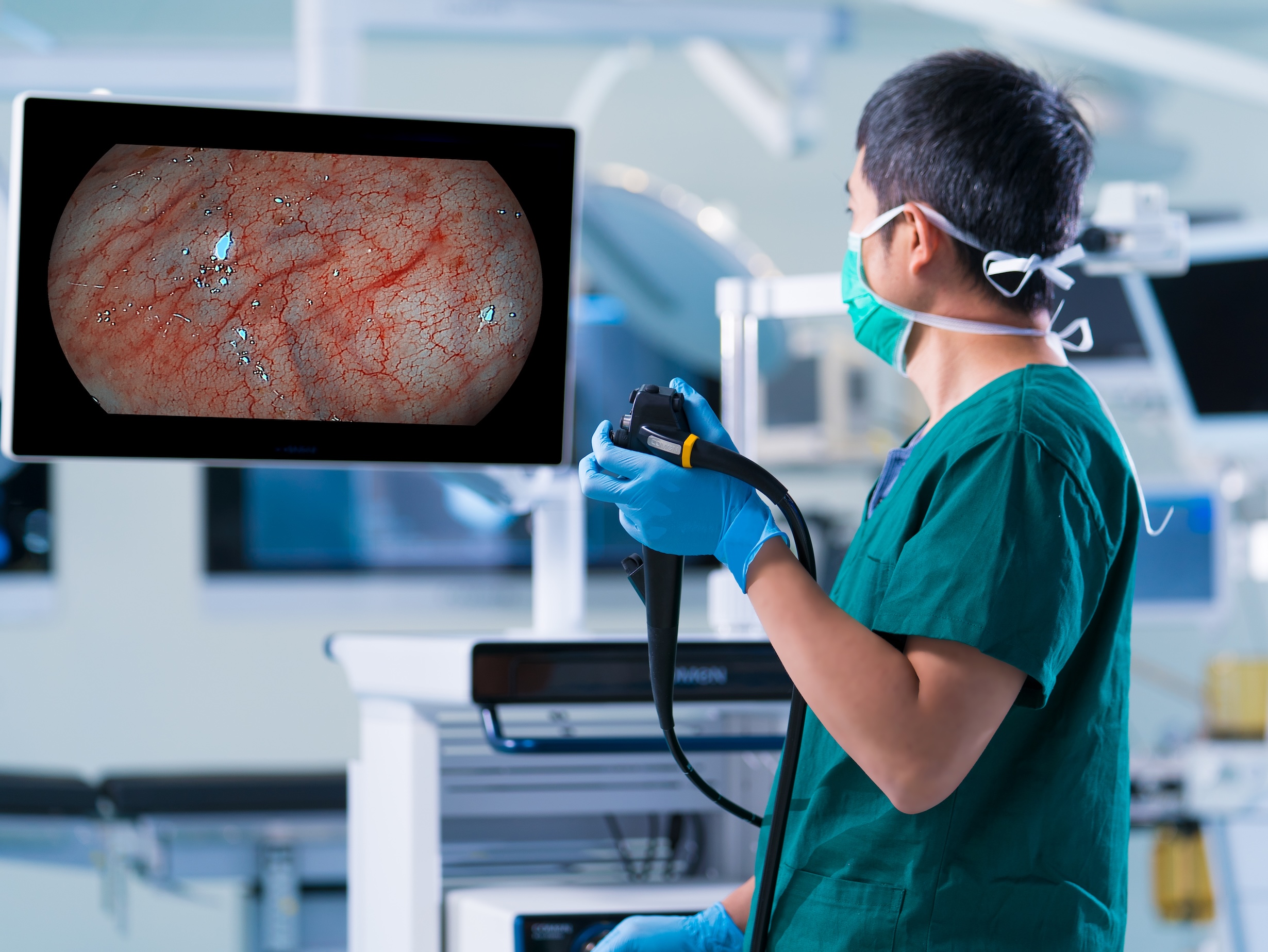
I’ll close with three concrete evaluation metrics I use when advising buyers: 1) Frame integrity rate under sustained capture (target ≥99.5% over 30 minutes), 2) Color fidelity deviation between capture and monitor (ΔE ≤ 3 in standard test targets), 3) Mean time to recover from a channel or illumination fault (target ≤5 minutes). Measure these; weigh them against total cost of ownership — not just sticker price. We ran these tests in a December 2022 procurement and the winning vendor reduced average case time by 9% within two months.
Choosing well requires discipline, testing, and a refusal to accept temporary fixes — I’ve lived the procurement headaches and the OR relief when the right systems arrive. For practical sourcing and implementation, consider vendors who back performance with real lab data and clear SLAs — like COMEN.